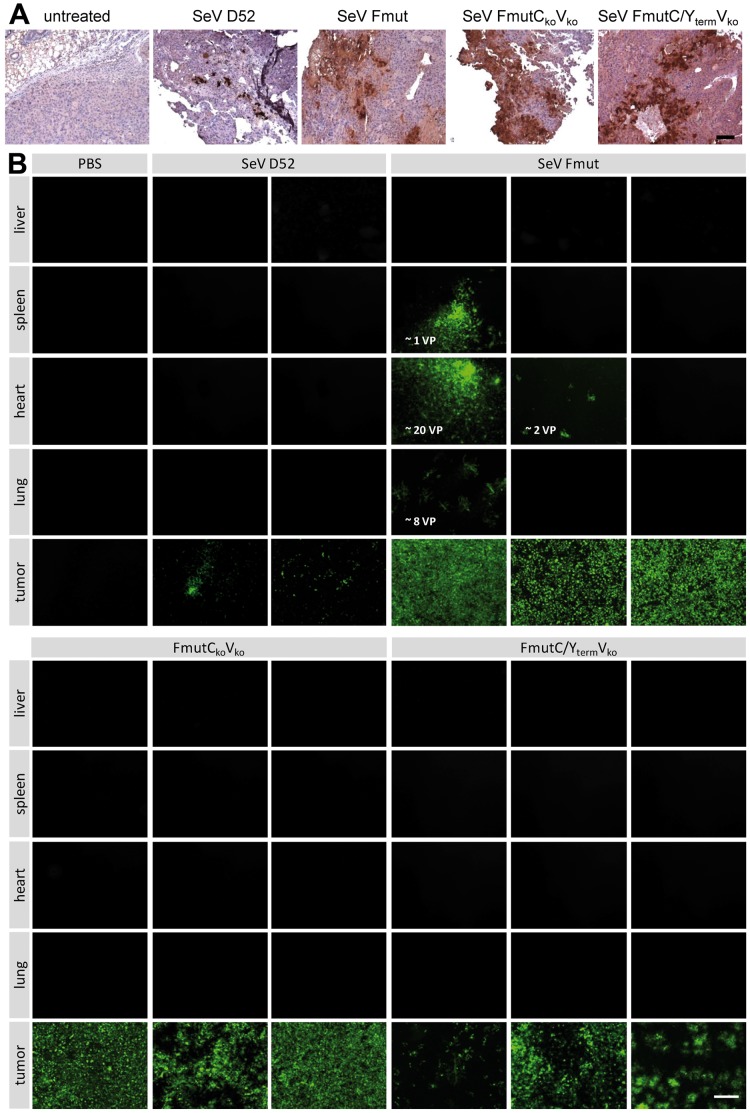
Figure 8. Intratumoral spread of recombinant SeV particles in vivo in a PLC/PRF/5 hepatoma xenograft model.
Recombinant SeV variants (SeV D52, SeV Fmut, SeV FmutCkoVko, SeV FmutC/YtermVko, 1×10⁷ TCID50/100 µl) were injected intratumorally in tumors of a PLC/PRF/5 hepatoma xenograft mouse model. (A) 48 h post injection the mice were sacrificed, tumors were removed and one quarter of each tumor was fixed and embedded in paraffin. Virus spread was investigated applying an anti-GFP antibody for immunohistochemistry analysis (brown color). Bar represents 100 µm. (B) Indicator cultures (Vero cells) were infected with lysates from frozen tissue sections (one quarter of tumor, liver, spleen, heart, lung, each). Early primary infections (24-72 hpi) were observed and single infected cells were counted as initial virus particles (VP). Fluorescence microscopy pictures of the infected Vero cells were taken 72 hpi. Shown are representative picture for each animal. Bar represents 400 µm.

