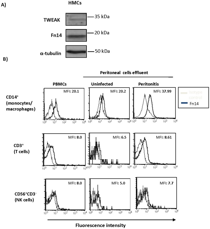
Figure 2. Fn14 is expressed by cultured human mesothelial cells and by leukocytes in peritoneal effluents from PD patients.
A) Cultured human mesothelial cells (HMC) express Fn14 and TWEAK as assessed by western blot. Representative images of three different experiments. B) Fn14 expression was analyzed by flow cytometry in peripheral blood and peritoneal effluent leucocytes. Cells were stained with Fn14-PE and monocyte/macrophages, T cells and NK cells were identified with anti-CD14, anti-CD3 and anti-CD56 antibodies respectively within the appropriate gates according to FSC and SSC parameters. Controls for the technique were stained with isotype immunoglubulin. Peripheral blood mononuclear cells (PBMCs) were used as positive controls for leukocyte population markers. Numbers within histograms indicate mean fluorescence intensity (MFI) for Fn14 staining. Note increased Fn14 expression mainly in peritoneal macrophages from patients with peritonitis.

