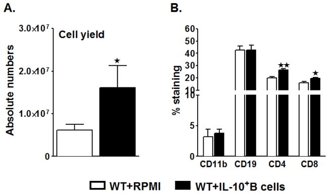
Figure 2. B-cell-sufficient (WT) mice transferred with IL-10-GFP+ B-cells before MCAO-induction have significantly reduced splenic atrophy.

Ninety six hours after MCAO, mononuclear cells were isolated from spleens of RPMI or IL-10-GFP+ B-cell recipient WT mice and analyzed for: A. Total cell count via hemocytometer. Values represent mean numbers (±SEM) of indicated cell subsets from 16–17 mice in each group, from at least 5 separate experiments; B. Comparison of CD11b+ monocytes, CD19+ B-cells, CD4+ and CD8+ T-cell populations. Values represent mean numbers (±SEM) of indicated cell subsets, gated on live leukocytes (by PI exclusion), from 6–7 mice of each group, from at least 2 separate experiments. Statistical analysis was performed with Student’s t-test to compare between RPMI and IL-10-GFP+ B-cell recipient mice. Significant differences between sample means are indicated (*p≤0.05).
