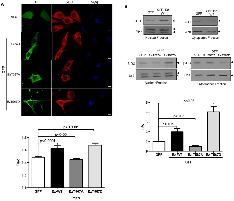
Figure 2. Overexpression of active ezrin facilitates nuclear translocation of β-DG.
A. C2C12 myoblasts cultured on glass coverslips were transfected to express ezrin-GFP (Ez) fusion proteins (either wild type, WT, or the mutated variants T567D and Ez-T567A) or GFP alone. Cells were fixed and stained 24 h post-transfection with a polyclonal anti-β-DG antibody (JAF) and TRITC-conjugated secondary antibody, with nuclei stained using DAPI (blue). Cells were imaged by CLSM, with typical single Z-sections shown (scale bar is 10 µm). Quantitative analysis for the nuclear to cytoplasmic ratio (Fn/c) of β-DG was performed (bottom panel) using the Image J software, as described in Material and Methods. Results represent the mean +/– SD (n > 50 cells) from a series of three separate experiments, with significant differences between cells expressing GFP alone and cells expressing the different GFP-tagged ezrin variants determined by Student t-test. B. Cytoplasmic and nuclear extracts obtained from cells transfected to express the above constructs were separated by SDS-PAGE and subjected to Western analysis for β-DG. Membranes were stripped and reprobed for Sp3 and calnexin (Clnx); loading controls for nuclear and cytoplasmic extracts respectively. Densitometric analysis of autoradiograms was performed, and the nuclear/cytoplasmic ratio (n/c) for β-DG obtained by dividing the relative levels of β-DG in the nuclear extracts with those obtained in the corresponding cytoplasm extracts (bottom panel). Results represent the mean +/– SD for 3 separate experiments, with significant differences between cells expressing GFP alone and those expressing the different GFP-tagged ezrin variants determined by Student t-test.

