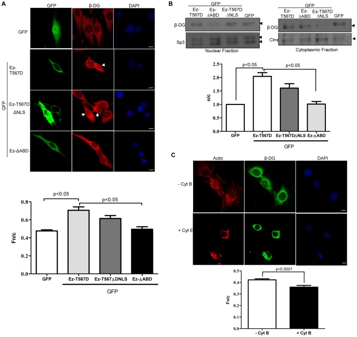
Figure 4. Ezrin-mediated cytoskeleton reorganization facilitates nuclear translocation of β-DG.
A. C2C12 myoblasts grown on coverslips were transfected to express GFP-tagged-EzT567D (active ezrin), -EzT567ΔNLS (active ezrin carrying a deletion of the NLS), or -EzΔABD (ezrin variant with a deletion of the actin-binding domain), or GFP alone. Transfected cells were immunostained with anti-β-DG primary and TRITC-conjugated secondary antibodies and counterstained with DAPI (nuclei), prior to CLSM, with typical single Z-sections shown (scale bar is 10 µm). Images were analysed to determine the Fn/c for β-DG (bottom), as per Figure 2. Results represent the mean +/– SD (n > 50) from three separate experiments, with significant differences in the Fn/c of β-DG between cells expressing GFP alone and those expressing GFP-Ez-T567D, and between cells expressing GFP-Ez-T567D- and GFP-EzΔABD, as denoted by the p values. B. Cytoplasmic and nuclear extracts from transfected cells with the above constructs were analyzed by SDS/Western blotting using anti-β-DG antibodies. Sp3 and calnexin (Clnx) were immunodetected as loading controls for nuclear and cytoplasmic fractions respectively. Densitometric analysis was performed to obtain the n/c ratio of β-DG for the different transfected cultures (right panel), as per Figure 2. Results represent the mean +/- SD for 3 separate experiments, with significant differences between cells expressing GFP alone and those expressing GFP-Ez-T567D, as well as between cell expressing GFP-Ez-T567D and those expressing GFP-EzΔABD, as denoted by the p values. C. C2C12 myoblasts seeded in coverslips were treated without (control) or with 6 µM cytochalasin B (Cyt B) for 1 h. Treated cells were fixed and double-stained with anti-β-DG primary antibody and fluorescein-conjugated secondary antibody, and with TRITC-phalloidin to visualize effects on the actin-based cytoskeleton network. Nuclei were counterstained with DAPI. Samples were imaged and subjected to image analysis as described in Figure 2. Results represent the mean +/- SD for three separate experiments (n> 50), with p values determined by Student t-test denoting significant differences between control and Cyt B-treated cells (bottom panel).

