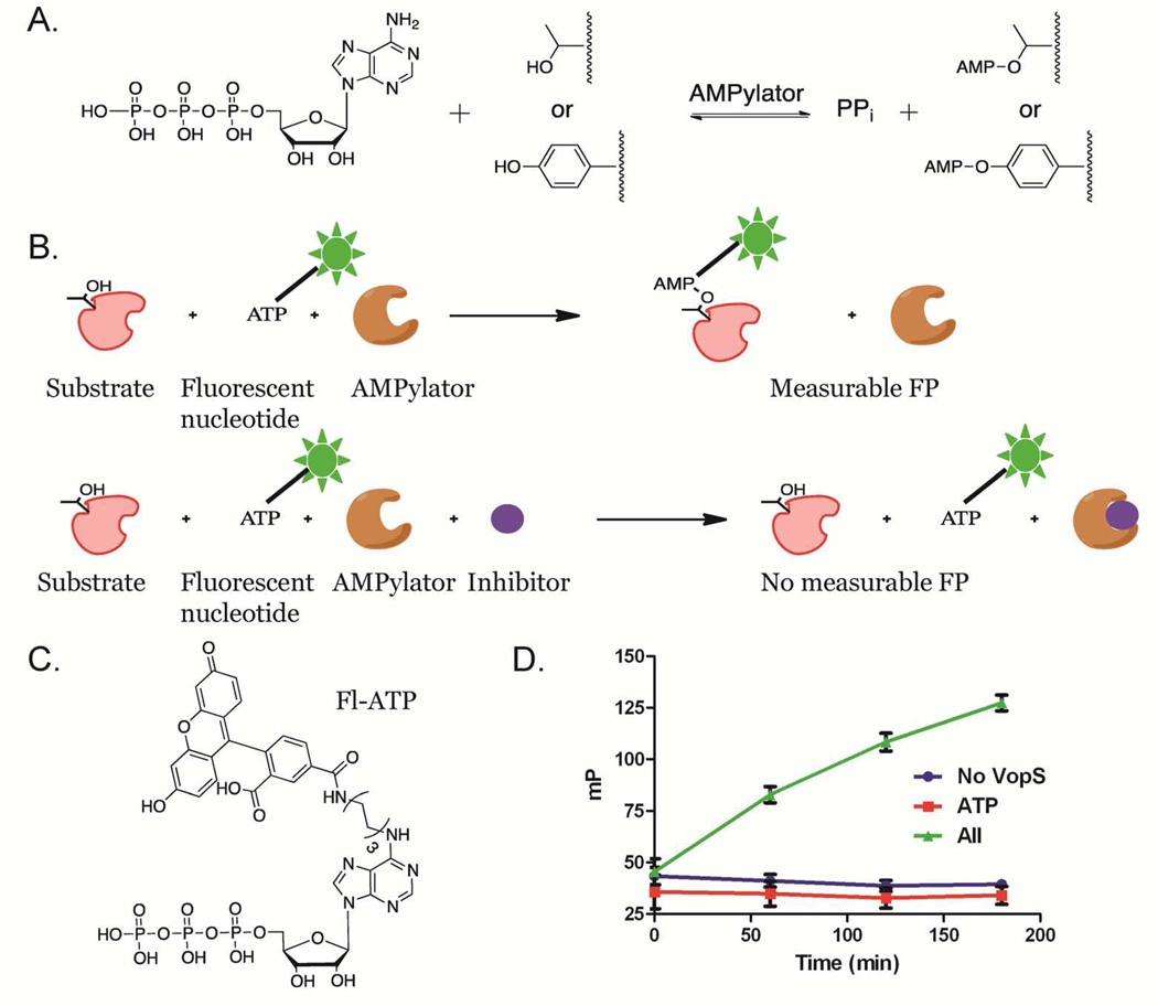
Figure 1.
VopS reaction and assay design. a) AMPylators transfer AMP to side chain threonines or tyrosines. b) FP assay design. A time dependent increase in FP signal occurs as VopS transfers Fl-AMP to Cdc42. In the presence of an inhibitor, however, no Fl-AMP is transferred and the FP signal is diminished. c) Structure of Fl-ATP. d) Substrates (Cdc42 and Fl-ATP) were added to 16 replicates of VopS, VopS with 0.4 mM ATP or VopS Screening Buffer in a 384-well plate. FP was measured as a function of time.

