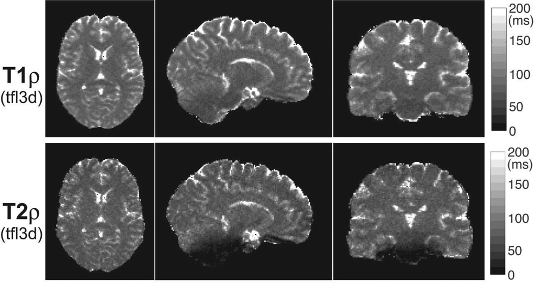
Figure 11.
3D T1ρ and T2ρ maps of the brain acquired in a volunteer. Five preparation periods of 0, 16, 32, 48 and 64 ms consisting of 0, 4, 8, 12 and 16 GOIA-W(16,4) pulses were acquired with 3D turbo FLASH at 1.33 mm isotropic resolution in 5:40 min. T1ρ map shows good uniformity in all three orthogonal cross-sections. T1ρ map shows good uniformity in all three orthogonal cross-sections. T2ρ map shows signal loss towards the base of the skull, predominantly in vermis and pons as seen in the sagittal and coronal views. Good contrast can be seen between gray and white matter, and subcortical structures.

