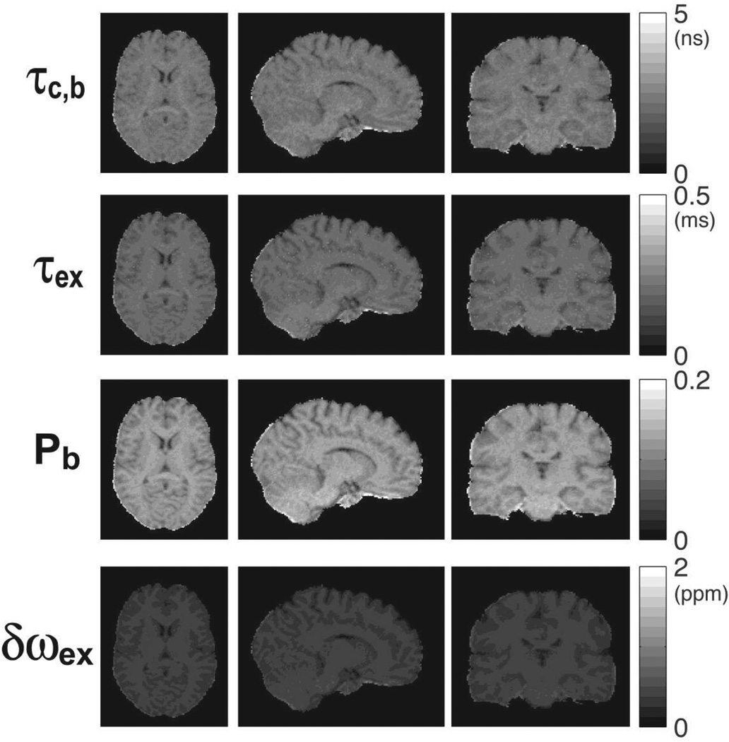
Figure 13.
3D parametric maps of water molecular dynamics in the brain of a human volunteer. The maps were obtained by fitting the 2SX/FXL model to rotating frame relaxation data from Fig. 11. Maps are uniform and without artifacts in all three orthogonal sections. Good contrast can be seen in all maps between gray and white matter, and subcortical structures.

