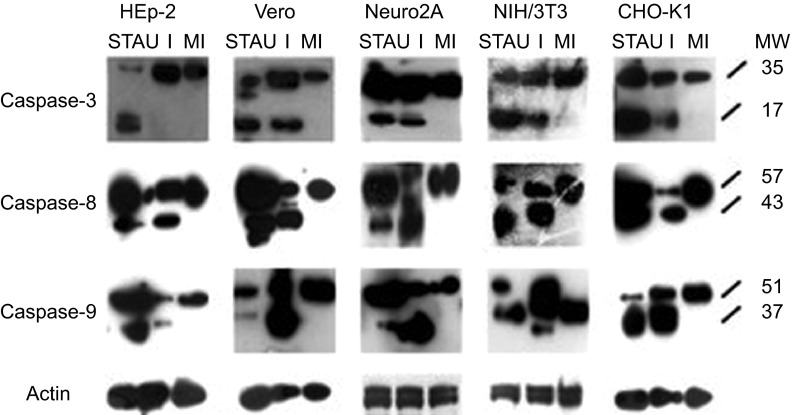
Figure 5.
Western blots of cell lysates derived from SAFV-infected HEp-2, Vero, Neuro2A, NIH/3T3 and CHO-K1 cell lines harvested at mid CPE in comparison with respective mock-infected control and STAU-treated control cells. Membranes were stained with respective antibodies against caspases-3, -8, -9 and actin. The intensity of actin staining served as the protein loading control. Lanes with I denote infected cell lysates, lanes with MI denote mock-infected control cell lysates, lanes with STAU denote STAU-treated control cell lysates and lanes with MW indicate the protein molecular mass in kDa. Caspases-3, -8 and -9 were cleaved to their respective active forms (p17, p43 and p37) in SAFV-infected Vero, Neuro2A, NIH/3T3, CHO-K1 cells and all STAU-treated cells. Only caspases-8 and -9 were cleaved to their active forms in SAFV-infected HEp-2 cells. The Western blotting results shown here are representative one of the three replicate experiments.

