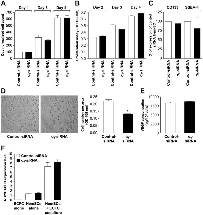
Figure 4. Effect of α6-integrin knock-down on proliferation, stem cell antigen expression, adhesion and differentiation of HemSC.
A- Proliferation of siRNA transfected HemSC cultured in EBM-2/20% FBS over 4 days evaluated by counting cells.
B- Proliferation of transfected HemSC cultured in EBM-2/20% over 4 days evaluated by measuring cellular phosphatase activity.
C- Expression of stem cell antigens CD133 and SSEA-4 inHemSC determined by flow cytometry 3 days after siRNA transfection with control or α6-integrin siRNA.
D- Adhesion assay: cells allowed adhering to laminin-coated wells for 20 minutes. Number of adherent cells determined by the p-NPP colorimetric assay (right panel: original magnification, ×10).
E- VEGF-A secreted by siRNA transfected HemSC measured after 3 days by ELISA on HemSC supernatant.
F- siRNA transfected HemSC co-cultured with cord blood ECFC for 5 days and separated into endothelial and non-endothelial cells using anti-CD31-coated magnetic beads. CD31-negative (non-endothelial) fraction analyzed by RT-qPCR for pericytic marker NG2 (neural glial antigen-2) and results compared with HemSC and cord blood ECFC cultured alone. No difference observed between control-siRNA and α6-integrin siRNAHemSC.

