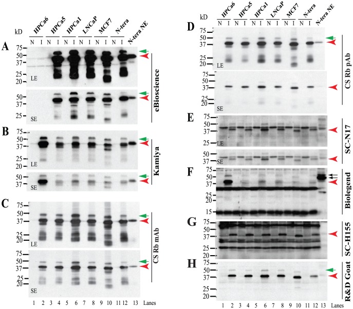
Figure 6. Reactivity of rNanogP8 and rNanog proteins towards 8 anti-Nanog Abs.
WB analysis using 8 anti-Nanog Abs (A-H). Cell types from which the initial cDNAs were cloned are indicated on top. Individual Abs are indicated on the right and M.W on the left. For some Abs, both a long (LE) and short (SE) exposures were shown. N: non-induced; I: induced by IPTG (see Methods). The red arrowheads in each panel indicate the 42 kD major Nanog protein and green arrows point to minor upper bands. In panel F, the two arrows point to the ∼48/54 kD doublets recognized by the BioLegend Rb pAb.

