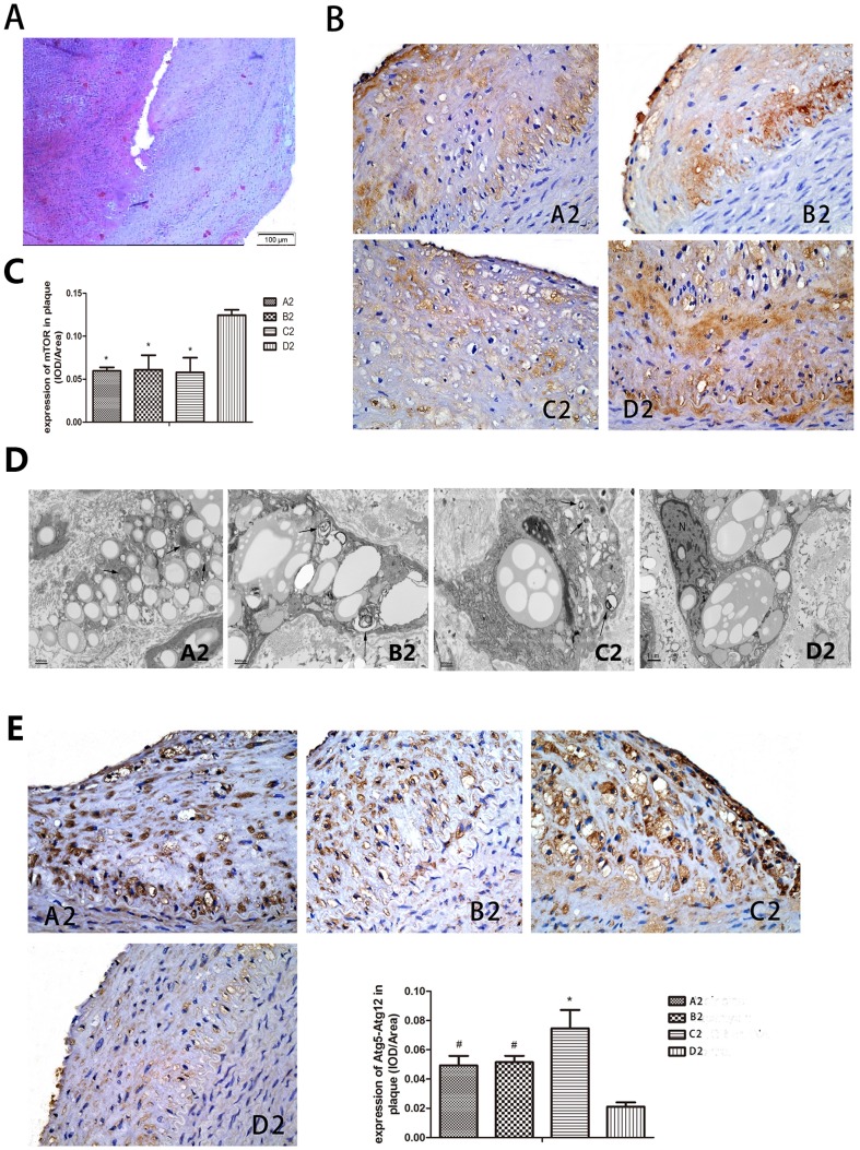
Figure 3. Effect of Pharmacological triggers in the experiment showed the rupture of plaque incidence higher in control group.
IH staining and transmission electron microscope showed mTOR decreased and autophagy increased in experimental group. Pharmacological triggers were done by 0.15·kg−1 of Chinese Russell's viper venom injecting intraperitoneally, 30 min later, 0.02 mg·kg−1 histamine was injected intravenously. (A) Haematoxylin and eosin staining of the cross section of the abdominal aorta in a rabbit of group D2 showing plaque rupture and thrombosis. (B, C) IH staining showed mTOR decreased obviously in group A2, B2 and C2 than D2. (D) Transmission electron microscope analysis showed macrophage autophagy was enhanced in group A2, B2 and C2 compared to group D2. Arrows in picture B2, C2 and d2 represent vacuoles with cytoplasmic inclusions and myeline figure. The “N” represents the nuclear. (E) IH analysis of Atg5-Atg12 conjugation found percentage of positive-stain cells was significantly increased in group A2, B2 and C2 in comparison to group D2. A2: triciribine; B2: rapamycin; C2: mTOR-siRNA; D2: control; * P<0.01 vs control, # P<0.05 vs control.

