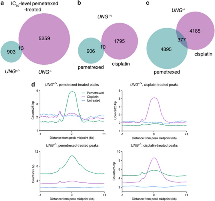
Figure 4.
γ-H2AX ChIP-seq in UNG+/+ and UNG−/− cells. Cells were treated with IC50-level pemetrexed (UNG+/+, 200 nM and UNG−/−, 25 nM) or with 20 μM cisplatin for 24 h and subsequently processed for ChIP-seq as described in the Materials and Methods section. Cisplatin was utilized as a control DNA-damaging agent that forms DNA double-strand breaks. (a–c) Venn diagrams of the ChIP-seq peaks generated comparing signal overlap for pemetrexed treated UNG+/+ and UNG−/− cells (a); pemetrexed treated and cisplatin treated UNG+/+ cells (b) and pemetrexed-treated and cisplatin-treated UNG−/− cells (c). (d) Midpoint coordinates for each γ-H2AX peak were used to obtained signals ±1 kb for the coordinate (within a 25-bp window). The average signal in each window for each peak shown for UNG+/+ pemetrexed treated (top left); UNG+/+ cisplatin-treated (top right); UNG−/− pemetrexed-treated (bottom left) and UNG−/− cisplatin-treated cells (bottom right)

