Figure 6.
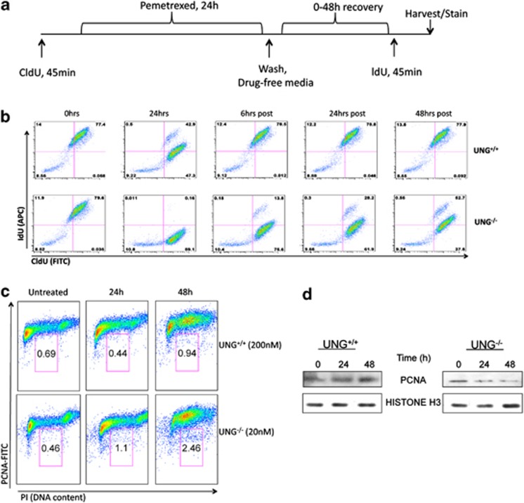
Intracellular replication fork instability in UNG−/− cells. (a) Schematic for CldU and IdU pulse labeling of pemetrexed-treated cells. Briefly, cells were incubated for 45 min with CldU (50 μM) prior to treatment with IC50-level pemetrexed (UNG+/+, 200 nM, UNG−/− 25 nM) for 24 h. Cells were then allowed to recover in drug-free media for 0–48 h prior to incubation for 45 min with IdU (50 μM). Cells were then stained with fluorescent antibodies that recognize CldU (rat-antiBrdU-FITC) and IdU (mouse-antiBrdU-APC). Representative data from flow cytometry sorting of CldU/IdU-labeled cells is shown in (b) where the x axis is CldU (FITC) and the y axis is IdU (APC). (c) Cells were treated with IC50-level pemetrexed for 24 and 48 h and subsequently stained with PI (x axis) to and FITC-labeled PCNA antibody (y axis). (d) Cells were treated as in (c) and subsequently incubated in 1% formaldehyde for crosslinking and chromatin extraction for western blots of chromatin-bound PCNA and histone H3 (loading control)

