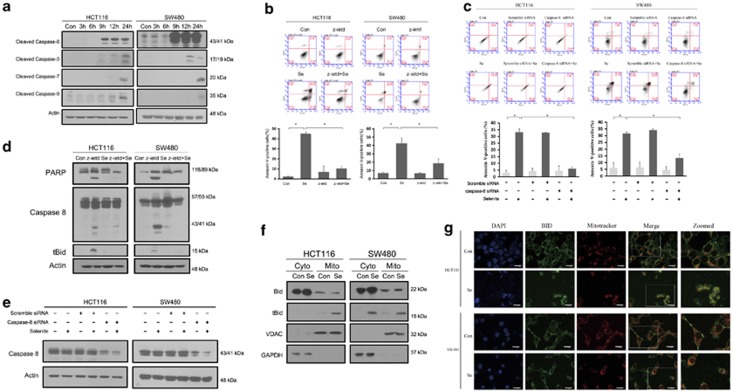
Figure 1.
Selenite-induced apoptosis and activation of caspase-8. (a) Western blot analysis of activated caspases over time showing the response of HCT116 and SW480 cells to 10 μM selenite from 0 to 24 h. (b and c) HCT116 and SW480 cells were treated with the caspase-8 inhibitor z-IETD-fmk for 1 h before selenite treatment or were transfected with caspase-8 siRNA for 24 h before selenite treatment. The cells were then stained with Annexin V/PI to measure the ratio of apoptotic cells through FACS. (d and e) Cells were treated with z-IETD-fmk or transfected with siRNA-targeting caspase-8 and subjected to western blot analysis. (f) Selenite treatment caused the cleavage of Bid to form tBid and subsequent translocation of tBid from the cytoplasm to the mitochondria. Mitochondria were isolated after selenite treatment and immunoblotted for Bid and tBid. VDAC and GAPDH were used as mitochondrial and cytoplasmic markers, respectively. (g) Confocal analysis of Bid localization in selenite-treated HCT116 and SW480 CRC cells. Mitotracker (red) solution was added to the medium 1 h before harvesting cells for mitochondrial staining. The cells were collected and incubated with an antibody to detect both Bid and tBid and then stained with a FITC-conjugated secondary antibody (green). The nuclei are shown as blue signals. Scale bar, 20 μm. The statistical graphs are presented as the mean±S.D. of three independent experiments; *P<0.05

