Figure 3.
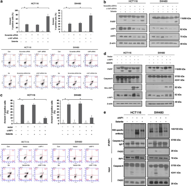
Selenite-induced degradation of cIAPs facilitated apoptosis by promoting DISC formation. (a and b) After cIAP silencing using an siRNA mixture containing sequences targeted to cIAP1 and cIAP2 for 24 h, HCT116 and SW480 CRC cells were treated with 10 μM selenite and subjected to FACS and western blot analysis. (c and d) HCT116 and SW480 cells were transfected with a plasmid expressing cIAP1 before selenite treatment and were then subjected to FACS and western blot analysis. (e) Coimmunoprecipitation using a RIP1 antibody was performed after transfection of CRC cells with the cIAP1 plasmid, followed by incubation with K63 polyubiquitin chain-specific antibody, caspase-8 antibody and FADD antibody

