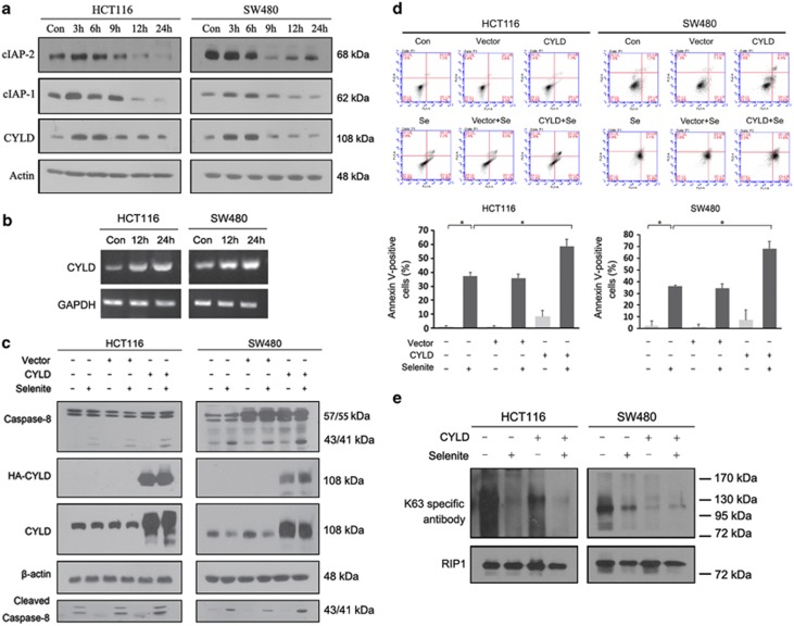
Figure 4.
CYLD was responsible for apoptosis induction via RIP1. (a) Western blot analysis of cIAPs and CYLD protein levels during a 0–24 h time course in HCT116 and SW480 cells treated with 10 μM selenite. (b) Selenite increased the level of CYLD transcription in HCT116 and SW480 cells. The cells were treated with selenite for the indicated time periods followed by reverse transcription and PCR. (c and d) HCT116 and SW480 cells were transfected with the CYLD expression plasmid. Twenty-four hours post-transfection, the cells were treated with selenite (10 μM) for 24 h, and the proteins were then extracted and subjected to western blot and FACS analysis. (e) RIP1 was coimmunoprecipitated from HCT116 and SW480 cells transfected with the CYLD plasmid and treated with 10 μM selenite 24 h after transfection. The resulting pellet was analysed by western blotting using antibodies against K63 polyubiquitin chains

