Figure 3.
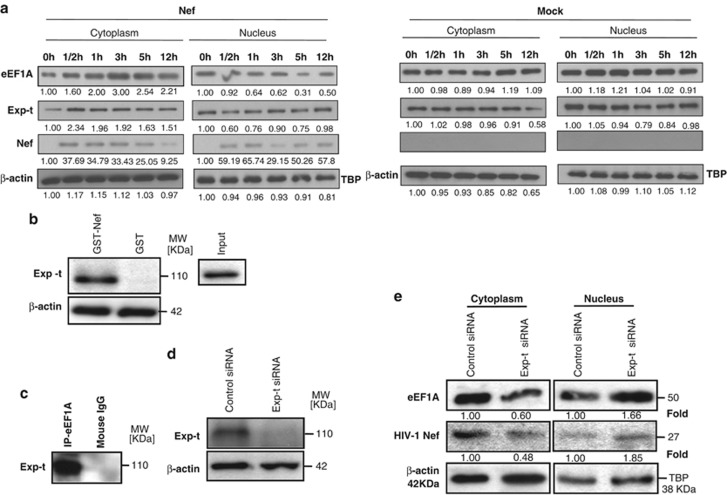
Nuclear–cytoplasmic relocalization of eEF1A/rNef occurs in MDMs treated with rNef and depends on Exp-t. (a) Kinetics of eEF1A/rNef/Exp-t in nuclear and cytoplasmic compartments of MDMs. Nuclear and cytoplasmic extracts of MDMs treated with rNef (100 ng/ml) were prepared and the expression of eEF1A, Exp-t and rNef were assessed in both cellular compartments up to 12 h post treatment using western blotting. Similarly, nuclear and cytoplasmic extracts of untreated MDMs (mock) were prepared and tested for the expression of eEF1A, Exp-t and rNef. β-actin and TBP were used as loading controls. Results are representative of three independent experiments. (b) Using wild-type GST–Nef constructs, the binding of Exp-t present in MDM lysates was assessed in GST pull-down assays. Input corresponds to 10% of the material used for pull-down. Results are representative of two independent experiments. (c) eEF1A interacts with Exp-t in MDM lysates. Total MDM extracts were prepared and the eEF1A/Exp-t interaction assessed by immunoprecipitation with an anti-eEF1A antibody and western blotting with an anti-Exp-t monoclonal antibody. Results are representative of three independent experiments. (d) Knockdown of the Exp-t protein by siRNA in MDMs. MDM cultures were transfected with a scrambled control or Exp-t siRNA and total cellular extracts were prepared 48 h post transfection. Protein expression was analyzed by western blot. β-actin was used as a loading control. (e) Effect of Exp-t siRNA on nuclear–cytoplasmic transport of eEF1A and rNef in MDMs. MDM cultures were transfected with a scrambled control or Exp-t siRNA for 48 h before treatment with rNef (100 ng/ml) for 3 h. Nuclear and cytoplasmic extracts were prepared and analyzed by western blot using anti-Nef and anti-eEF1A antibodies. β-actin and TBP were used as loading controls. Results are representative of two independent experiments. Protein levels of eEF1A and Nef after siRNA transfection were quantified by densitometry using ImageJ 1.40 software (protein levels in cells transfected with scrambled siRNAs were arbitrarily established at 1)

