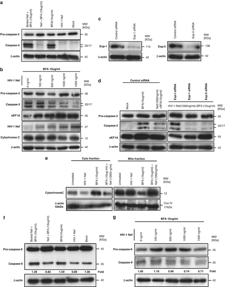
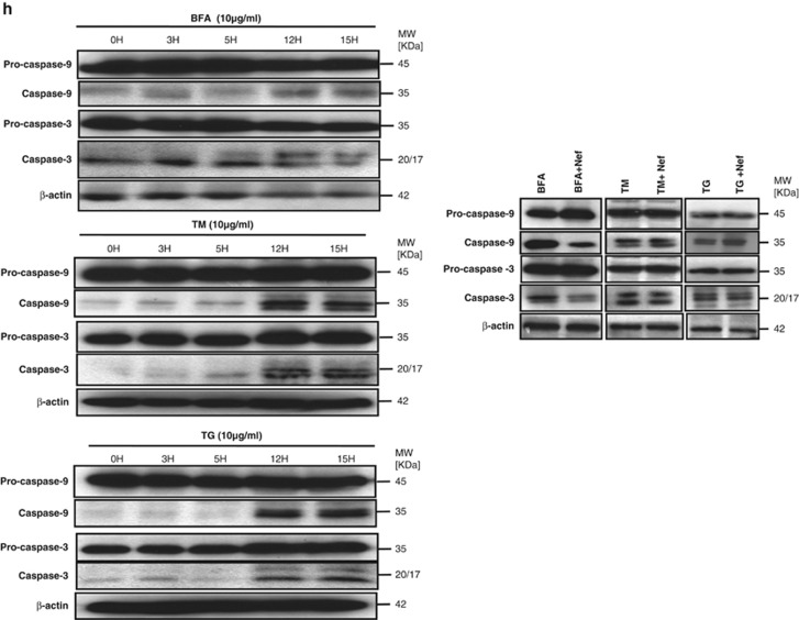
Figure 5.
rNef-mediated cytoplasmic accumulation of eEF1A in BFA-treated MDMs inhibits caspase activation and decreases the cytoplasmic release of cytochrome c. (a) Inhibition of caspase-3 activation in BFA-stimulated MDMs treated with rNef (1000 ng/ml). (b) Inhibition of caspase-3 activation in BFA-stimulated MDMs treated with rNef is dose-dependent. Mitochondrial cytochrome c release in BFA-treated MDMs is blocked by rNef in a dose-dependent manner and positively correlates with cytoplasmic accumulation of eEF1A. (c) Knockdown of Exp-1 and Exp-5 proteins by siRNA in MDMs. MDM cultures were transfected with a scrambled control, Exp-1 siRNA, or Exp-5 siRNA. Total cellular extracts were prepared 48 h post transfection. Protein expression was analyzed by western blot. β-actin was used as a loading control. (d) Inhibition of caspase-3 activation in BFA-stimulated MDMs treated with rNef is dependent on Exp-t. (e) Mitochondrial cytochrome c release in BFA-treated MDMs is blocked by rNef. (f) Inhibition of caspase-9 activation in BFA-stimulated MDMs treated with rNef (1000 ng/ml). Protein levels of caspase-9 were quantified by densitometry using ImageJ 1.40 software (the level of caspase-9 in mock cells was arbitrarily established at 1). (g) Inhibition of caspase-9 activation in BFA-stimulated MDMs treated with rNef is dose-dependent. Protein levels of caspase-9 were quantified by densitometry using ImageJ 1.40 software (the level of caspase-9 in mock cells was arbitrarily established at 1). (h) Inhibition of caspase activation in BFA-stimulated MDMs treated with rNef, but neither in TM-stimulated MDMs nor in TG-stimulated MDMs treated with rNef. Left panel: time–course of caspase activation in BFA-stimulated MDMs, TM-stimulated MDMs or TG-stimulated MDMs. Right panel: inhibition of caspase activation in BFA-stimulated MDMs treated with rNef, but neither in TM-stimulated MDMs nor in TG-stimulated MDMs treated with rNef. MDM cultures were treated with BFA (10 μg/ml), TM (10 μg/ml), or TG (10 μg/ml) for 12 h in the presence of rNef (1 μg/ml), and caspase-3 and -9 activation was measured in total cellular lysates. Results are representative of data obtained in three independent experiments


