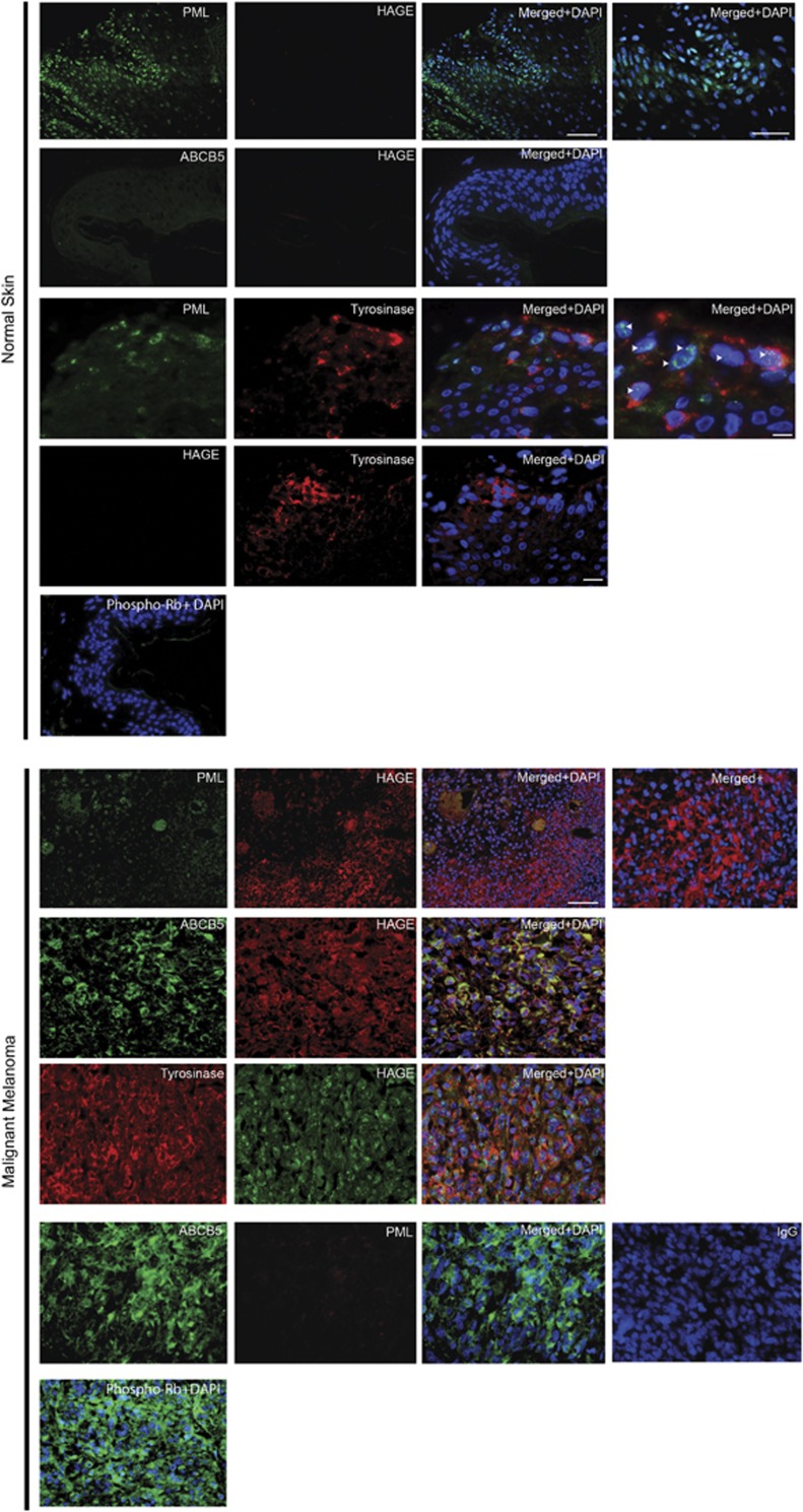
Figure 1.
Expression of PML in HAGE-positive and ABCB5-positive malignant melanoma. Top panel: representative image of immunohistochemistry with antibodies to PML (green) or ABCB5 (green) or Tyrosinase (red) or HAGE (red) or phospho-Rb (green) on normal skin sections. The arrows indicate PML-NBs in Tyrosine-positive cells. Lower panel: representative image of immunohistochemistry with antibody to PML (green or red), ABCB5 (green), Tyrosinase (red), HAGE (red) or phospho-Rb (green) in malignant melanoma tissue sections. The negative controls used were an IgG isotype from mouse and rabbit. The blue staining (DAPI) corresponds to the nucleus. Scale bars represent 100 μm (20 × ), 50 μm (40 × ), 20 μM (40 × )

