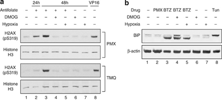
Figure 3.
Downstream effects of antifolates and BTZ under DMOG treatment demonstrated by western blot analysis. (a) Antifolate-induced H2AX phosphorylation. HeLa cells were exposed to antifolates (i.e., 1 μM PMX or TMQ) under normoxia for 24 or 48 h (lanes 2 and 3), 0.5 mM DMOG for 48 h with or without co-exposure to antifolates (lanes 4 and 6), hypoxia for 48 h with or without co-exposure to antifolates (lanes 5 and 7) and the DNA-damaging agent VP16 (40 μM for 20 min, lane 8). Total protein extracts were subjected to western blotting with a H2AX pS139 antibody. Equal loading was verified by reprobing with a histone H3 antibody. (b) BTZ-induced BiP overexpression. HeLa cells were exposed to 1 μM PMX for 24 h (lane 2), 80 nM BTZ under normoxia (lane 3), 0.5 mM DMOG for 24 h with or without co-exposure to 80 nM BTZ (lanes 4 and 6), hypoxia for 24 h with or without co-exposure to 80 nM BTZ (lanes 5 and 7, respectively) and the ER-stress-inducing agent tunicamycin (1 μg/ml for 24 h, lane 8). Cellular proteins were extracted using a hypotonic buffer and subjected to western blotting with a BiP antibody. Equal loading was verified by reprobing with a β-actin antibody

