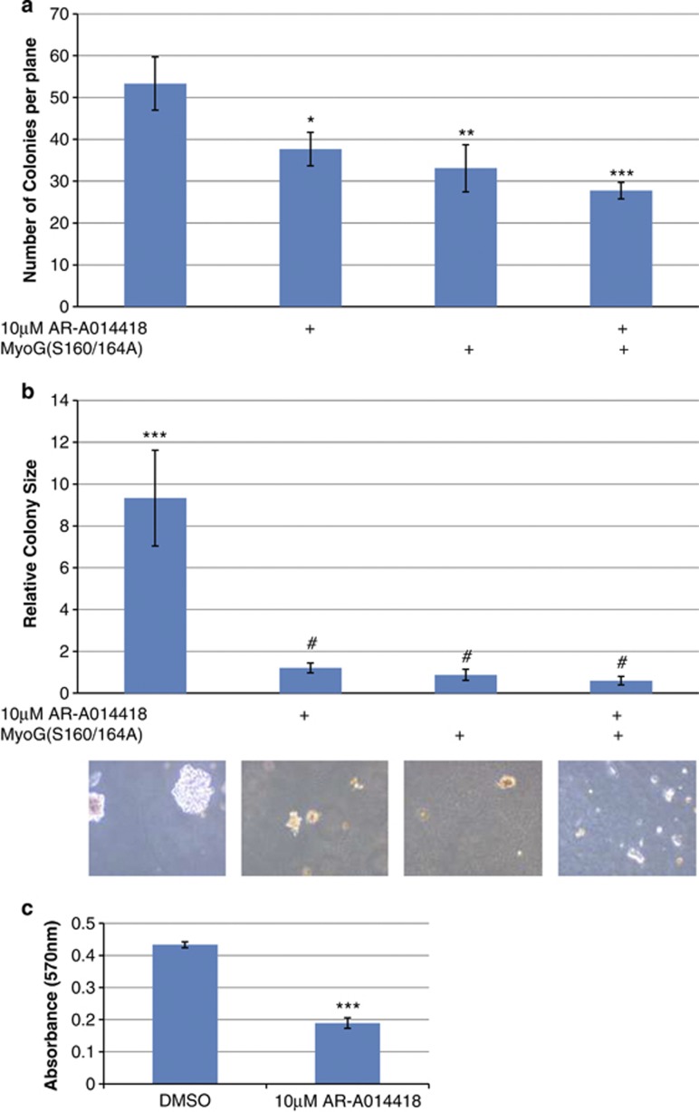
Figure 6.
Soft agarose colony formation and MTT cell proliferation assays: (a) Equal numbers of RH30 cells were seeded under different conditions as depicted, and allowed to form colonies for 21 days. On the 22nd day the colonies were stained with 0.005% Crystal Violet overnight. Colonies were counted at different planes (n=10) in four independent experiments done in triplicate. The total number of colonies was reduced by (i) 10 μM AR-A014418, P<0.05 (ii) Transient transfection of MYOGENIN containing the S160/164A neutralizing mutations, P<0.01 and (iii) both, P<0.001. Also see Supplementary Figure 1 for visual representation of the data. (b) We searched for the three largest colonies in each of the 12 plates for each condition and calculated the area using ImageJ software. The data revealed that the control colonies could grow up to 9 × bigger than any of the experimental conditions, P<0.001. (c) MTT cell proliferation assays were performed in PAX3/FOXO1A-expressing cells with and without 10 μM AR-A014418 treatment. The experiment revealed that GSK3β inhibition reduces cell proliferation by ∼2-fold, P<0.001. #ns, *P<0.05, **P<0.01, ***P<0.001

