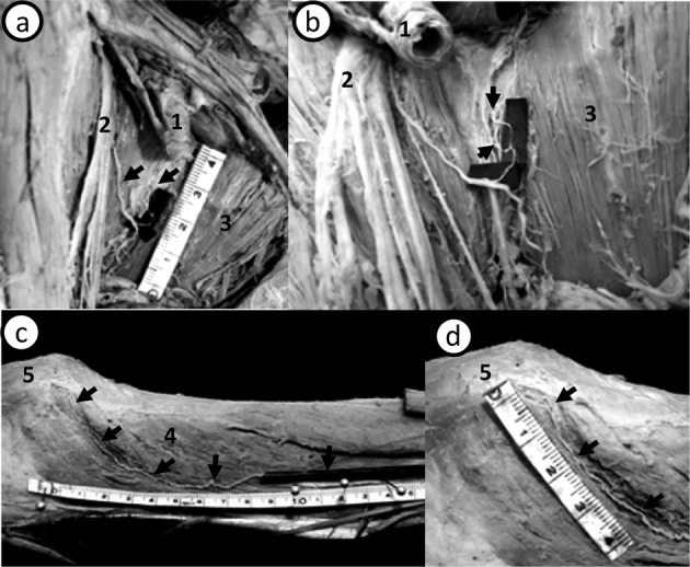
Fig. 1.

Small branches of the femoral nerve in the hip and knee joint. (a, b) The anteromedial aspect of the right hip joint. (c, d) The medial aspect of the right knee joint. 1: femoral artery. 2: femoral nerve. 3: pectineal muscle. 4: vastus medialis. 5: patella. In one cadaver, a small branch (arrow) that entered the anterior side of the iliofemoral ligament originated from the femoral nerve (a, b). In the same cadaver, one minor muscular branch that pierced the vastus medialis entered the joint capsule from the superior medial aspect of the patella (c, d). It seems that each branch is an articular branch of the femoral nerve innervating the hip and knee joints.
