Figure 5.
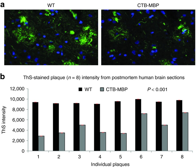
Ex vivoevaluation of amyloid plaque load in postmortem Alzheimer's disease (AD) human brain tissue sections. (a) Sections of the parietal cortex from human AD were incubated with 50 µg recombinant CTB-MBP protein partially purified from transgenic leaf extracts or 50 µg of partially purified wild type (WT) for 2 days and then stained with 0.02% thioflavin S (ThS) solution to visualize the central dense core of compact amyloid (green) and with DAPI (4′,6 diamidino-2-phenylindole) to identify cell nuclei (blue) by fluorescence microscopy. (Bar = 20 µm). (b) Quantification of the relative amounts of amyloid plaque load in the ThS-stained sections after incubation with the indicated proteins as described in (a). The mean plaque counts per section were determined with NIS Elements for Advanced Research. The values in the histogram represent the mean ± SD> n = 8; *P < 0.001 (single factor analysis of variance) relative to control. CTB, cholera toxin B subunit; MBP, myelin basic protein.

