
Fig. 4.
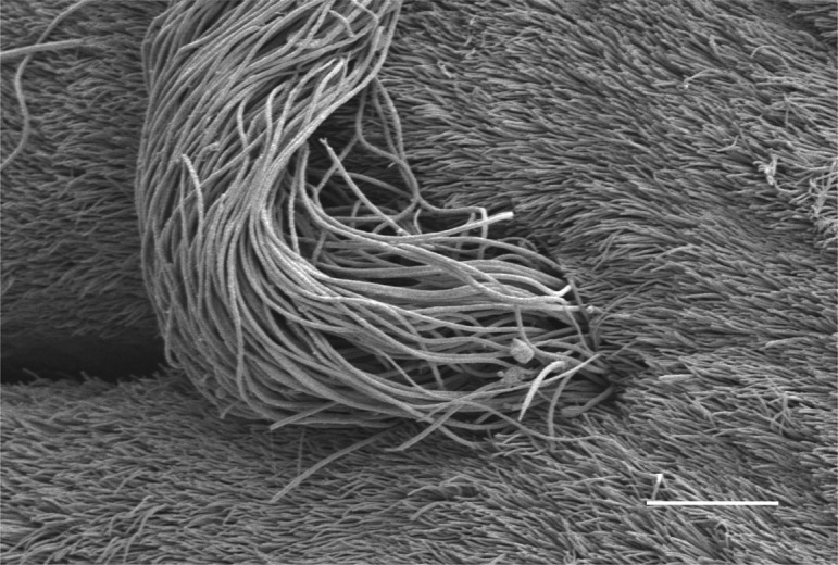
Ultrastructural observation of the UVJ surface treated with progesterone. After mating, the animals were injected with 0.8 μg/ml progesterone. The UVJ was isolated 1 h after the injection, and the SST entrance area was observed by scanning electron microscopy. A representative photograph from those obtained from three different birds is shown. Bar=10 μm.