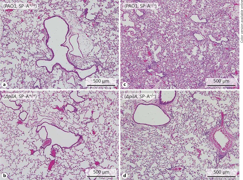
Fig. 2.
Histopathology of PA-infected lungs. SP-A+/+ and SP-A−/− mice were infected with PAO1 or ΔpilA as described in figure 1. Representative HE-stained lung sections from SP-A+/+ and SP-A−/− mice (n = 5) 18 h after intranasal instillation of PAO1 (a, c) and ΔpilA (b, d) bacteria.

