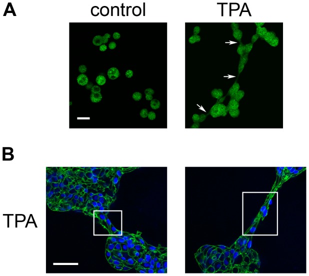
Figure 7. The bridges formed between acini in TPA-treated cultures contain actin.
On day 4, cultures of MCF10A acini were fed in the absence (control) or presence of 10 nM TPA (TPA), and fixed 21 hours later. A) Acini stained with phalloidin to detect actin (green) (10X); bar = 100 µM. Arrows indicate bridges. B) Acini stained with phalloidin and DAPI to detect nuclei (blue) (40X); bar = 50 µM. Boxes have been drawn around bridges. Representative images are shown.

