
Figure 5. Electrostatic potential surface of the allosteric binding pocket of HIV-1 IN CCD dimer in interaction with (A) BI-1001, (B) CX14442, and (C) LEDGF/p75.
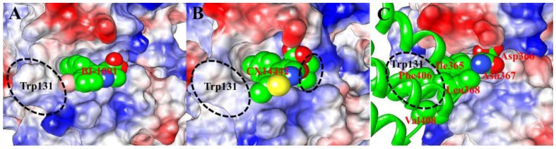
The positive charges are displayed in blue, negative charges are displayed in red, and neutral residues are displayed in white. Color intensity is proportional to the charge value. The BI-1001, CX14442 and side chain of the LEDGF/p75 key residues, whose carbon atoms are shown as green spheres and labeled as red. The residue Trp131 from monomer A of HIV-1 CCD dimer is also labeled (black).