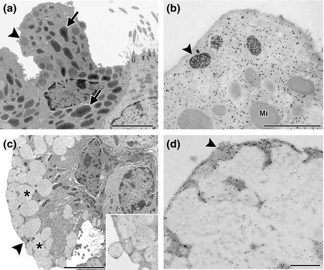
Figure 3.

Clara cells in chronic allergy. Electron microscopy (a and c) and immunoelectron microscopy for CC16 (b and d). In control mice, cells typically contained numerous mitochondria (arrow) and a few small secretory granules with round profiles and a moderate electron density (arrowhead) appearing under the plasma membrane (a). By immunogold labelling, CC16 can be observed in the cytoplasm, being very intense on some electron-dense apical secretory granules (arrowhead in b). Mi, mitochondria. In OVA challenged animals, cells appeared deeply modified, especially for the numerous large and electron-lucent fused secretory granules (*) in the whole apical cytoplasm, with abundant RER in the basal cytoplasm intermingled with some slim mitochondria (b). The large fused electron-lucent granules of these mucous transformed cells were positive for mucin (inset Figure 2b) and negative for CC16, with only a few positive small granules appearing under the plasma membrane (arrowhead) (d). Bar represents 1 μm.
