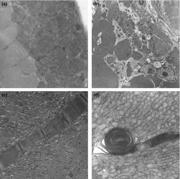
Figure 3.

Electron microscopic findings: (a) Aggregates of subsarcolemmal mitochondria in a 9 month old wild-type male mouse. (b) A necrotic muscle fibre undergoing phagocytosis in a 6 month old transgenic male mouse. (c,d) Tubular aggregates in a 6 month old male transgenic mouse and a spiral membranous body in one tubular aggregate (d). Magnification: (a) ×6610; (a) ×1300; (c) ×5200; (d) ×12600.
