
Figure 3.
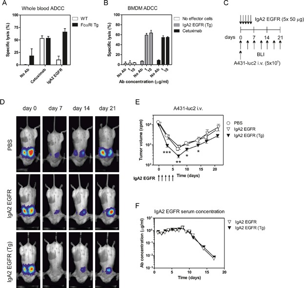
Anti-tumour activity of IgA2 EGFR in an intravenous xenograft model
- Specific lysis of A431-luc2 cells in vitro by G-CSF-primed mouse whole blood with 1 μg/mL EGFR antibodies in a 4 h 51Cr-release assay.
- Specific lysis of A431 cells by mouse bone marrow macrophages with EGFR antibodies in an overnight 51Cr-release assay.
- Scheme of A431-luc2 i.v. model. 5 × 10⁵ A431-luc2 cells were injected intravenously into FcαRI Tg SCID or WT SCID mice. Mice were injected i.p. with 50 μg IgA2 EGFR daily until Day 4. Tumour growth was monitored by serial BLI.
- Representative images from the BLI recordings of the A431-luc2 i.v. experiment.
- The curves represent the mean tumour volume (cpm) ± SEM for each measurement (seven mice/group, *p < 0.05, **p < 0.01, ***p < 0.001; Student's test, significance was assessed compared to the PBS group).
- Concentrations of IgA2 EGFR in serum during the A431-luc2 i.v. experiment. At each time point one mouse was bled from FcαRI Tg and WT groups.