
Figure 6.
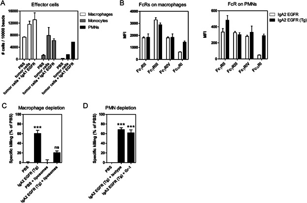
In vivo IgA2 EGFR activity is mediated by macrophages in a syngeneic peritoneal model using Ba/F3-EGFR cells
- Effector cells in the peritoneum in the Ba/F3-EGFR i.p. model. Effector cell types were identified by FACS and their relative number was determined compared to known amount of beads.
- Expression of FcRs on the different effector cell types in the peritoneal lavage during the Ba/F3-EGFR i.p. model in PBS-treated mice. Expression of mouse FcγRs and human FcαRI on macrophages (F4/80+) and PMNs (Ly6Ghigh) was analysed by FACS.
- Depletion of macrophages in the Ba/F3-EGFR i.p. model. Specific cytotoxicity of Ba/F3-EGFR cells in FcαRI Tg mice treated with 50 μg IgA2 EGFR or PBS. Macrophages were depleted prior to the experiment by injection of chlodronate liposomes (***p < 0.001, ANOVA, Bonferroni post test, four to five mice/group).
- Depletion of PMNs in the Ba/F3-EGFR i.p. model. Specific cytotoxicity of Ba/F3-EGFR cells in FcαRI Tg mice treated with 50 μg IgA2 EGFR or PBS. Prior to the experiment, mice were injected with 250 μg Gr-1 (Ly6G/C-specific) antibody, or with isotype control, two times intraperitoneally to deplete PMNs (***p < 0.001, ANOVA, Bonferroni post test, 9–10 mice/group).