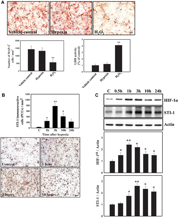
Figure 2.

- 4 h-hypoxia in PCCs was sublethal. Hypoxia was induced in a two-gas incubator (Jouan, Winchester, Virginia) equipped with an O2 probe to regulate N2 levels. Represented images (top panel) and summarized data (bottom left panel) for the number of viable MAP-2+ neurons were shown. Summarized data based on lactate dehydrogenase (LDH) assay was also shown (bottom right panel).
- The number of PCCs expressing STI-1 (B) and the total level of STI-1 expression in the cultures (C) were measured by means of immunocytochemistry and western blot, respectively. Hypoxia increased the number of PCCs expressing STI-1 (B) and the total protein level of HIF-1α and STI-1 in the cultures (C). Values are mean ± SEM. (*p < 0.05 and **p < 0.01 versus control). Scale bar, 50 µm.
