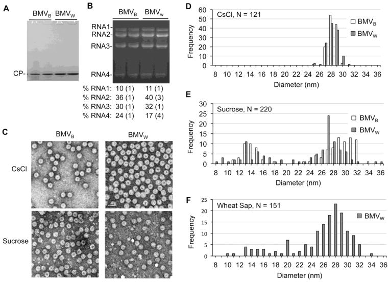
Fig. 7. Variation in the diameters of the BMV particles.
A) The CP is the most abundant protein from BMVW and BMVB purified by sucrose density cushions. 2 μg of total protein determined by optical density was loaded per lane in the SDS-PAGE. The gel was stained with Coomassie blue. B) Relative abundances of the virion RNAs from BMVW and BMVB enriched with sucrose cushions. RNAs was visualized by staining with ethidium bromide. The quantification of the relative amount of the RNAs were from three independent experiments. C) Negative-stained electron micrograph of BMVW and BMVB purified from CsCl gradients or the sucrose cushion. D) The size distribution of CsCl purified BMVW and BMVB. The outer diameters of the viral particles were measured from multiple micrographs to ensure random sampling. E) The size distribution of the BMVW and BMVB particles enriched through the sucrose cushion. G) The size distribution of BMVW for clarified wheat sap.

