
Fig. 8. BMV virions exhibit a range of densities in CsCl density gradients.
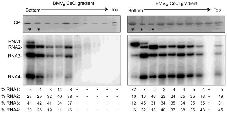
BMVW or BMVB enriched by sedimentation through a sucrose cushion were further centrifuged in a 45% cesium chloride gradient for 20 h at 50,000 g. Fractions were then collected following puncturing the bottom of the tube. The stars denote the fractions that appear in the opalescent white band. The upper images show the presence of the CP in each fraction. The SDS-PAGE gel was stained with Commassie blue. The lower images show the detection of viral RNAs by Northern blot. The quantification show the relative amount of the four viral RNAs in each fraction. Approximately equal amount of virions or viral RNAs from each fraction were used for the visualization, except for the fractions that contained significantly less viral RNAs.