Fig. 3.
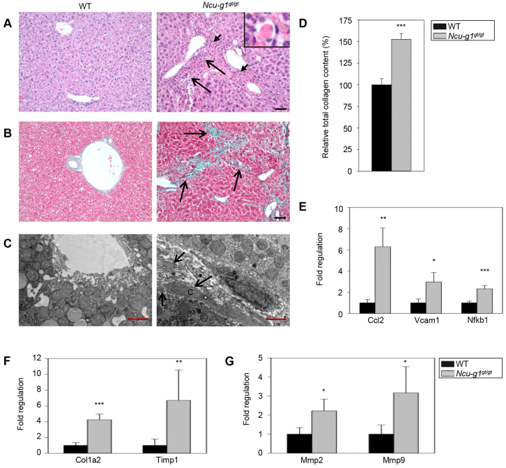
Inflammation, dying hepatocytes and collagen deposition in Ncu-g1gt/gt liver. (A) Hematoxylin and eosin staining of liver sections from 6-month-old wild-type (WT) and Ncu-g1gt/gt mice with a distorted tissue structure, massive infiltration of mononuclear leucocytes and granulocytes (long arrows), and acidophilic hepatocytes (short arrows, inset) in portal areas of Ncu-g1gt/gt mice. Scale bar: 50 μm. (B) Masson-Goldner’s trichrome staining of liver sections from WT and Ncu-g1gt/gt mice shows fibrosis in periportal regions of Ncu-g1gt/gt mice (arrows), where connective tissue stains green. Scale bar: 50 μm. (C) Representative transmission electron microscopy micrographs of perisinusoidal areas in liver sections from WT and Ncu-g1gt/gt mice. Massive deposition of fibrous collagen is present in the space of Disse of the Ncu-g1gt/gt liver (arrows) (C, collagen). Scale bars: 2 μm. (D) Relative total collagen content in WT and Ncu-g1gt/gt liver as measured by sirius-red colorimetric plate assay. (E) qPCR analyses show elevated expression of genes involved in leukocyte recruitment (Ccl2, Vcam1, Nfkb1), and (F,G) extracellular matrix remodeling (Col1a1, Timp1, Mmp2, Mmp9) in Ncu-g1gt/gt liver (n=4, *P<0.05, **P<0.005, ***P<0.001). Values are expressed as mean±s.e.m.

