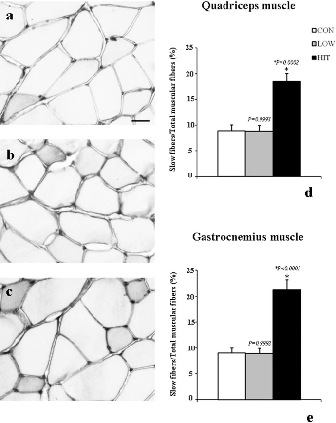
FIG. 1.
IMMUNOPOSITIVITY FOR SLOW SKELETAL MYOSIN IN THE QUADRICEPS AND GASTROCNEMIUS MUSCLES OF MOUSE AFTER EXERCISE TRAINING
Note: Representative immunoperoxidase stains for slow skeletal myosin in the quadriceps muscle from CON (a), LOW (b), and HIT mice (c) are shown. The immunopositivity for this protein is increased in mice after high-intensity exercise training (c), as confirmed by the histograms showing the percentage of slow fibres among total muscle fibres in the quadriceps muscle (d). The same result was observed in the gastrocnemius muscle (e).
*P < 0.001 versus CON and LOW
Scale bar= 33.48 µm

