
FIG. 2.
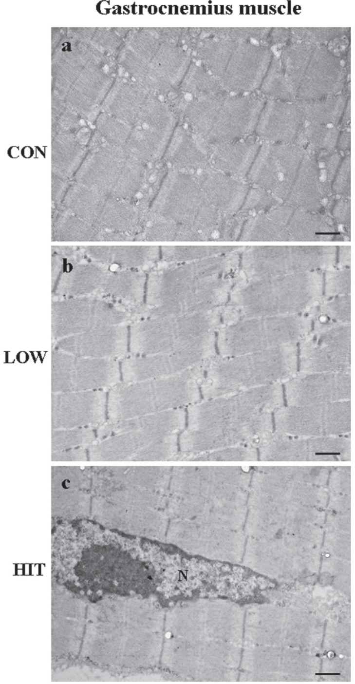
ULTRASTRUCTURAL ANALYSIS OF GASTROCNEMIUS MUSCLE OF MOUSE AFTER EXERCISE TRAINING
Note: Representative micrographs of gastrocnemius muscle from CON (a), LOW (b) and HIT mice (c). No ultrastructural alterations were detected in any of the mice groups. The myofibres are well formed, with repeating sections of dark and light bands.
In Fig. 2c the nucleus (N) of a myofibre.
Scale bars: a, b, c=0.62 m