
Figure 2.
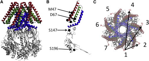
Crystal structure of MscS and spin-label locations. (A) 2oau closed crystal structure. Transmembrane helices 1, 2, and 3 are highlighted. Cytoplasmic domains are shown in light gray. (B) Monomer of MscS showing spin-labeling sites, which are shown as black spheres. (C) Incorporation of a single cysteine into an MscS monomer and subsequent spin labeling will lead to a heptamer with seven spin labels and three different spin-spin distances. To see this figure in color, go online.