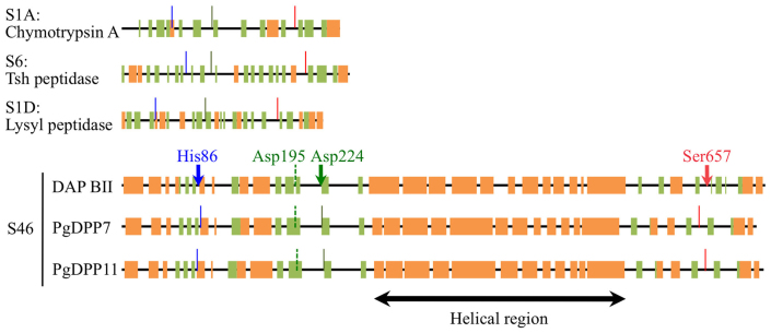
Figure 4. Secondary structure prediction of family S46 peptidases.
Secondary structures of family S46 peptidases as predicted by Jpred compared to those derived for chymotrypsin A from Bos taurus, Tsh peptidase from E. coli, and lysyl peptidase from Achromobacter lyticus. Regions that form a beta-sheet (green) or an alpha-helix (orange) are indicated by boxes, while solid lines and arrows show the His (blue), Asp (green) and Ser (red) residues of the catalytic triads. Arrows indicate the active residues identified in this study, while the broken green lines mark Asp residues that are part of the catalytic triad according to Ohara-Nemoto, Y. et al.22.

