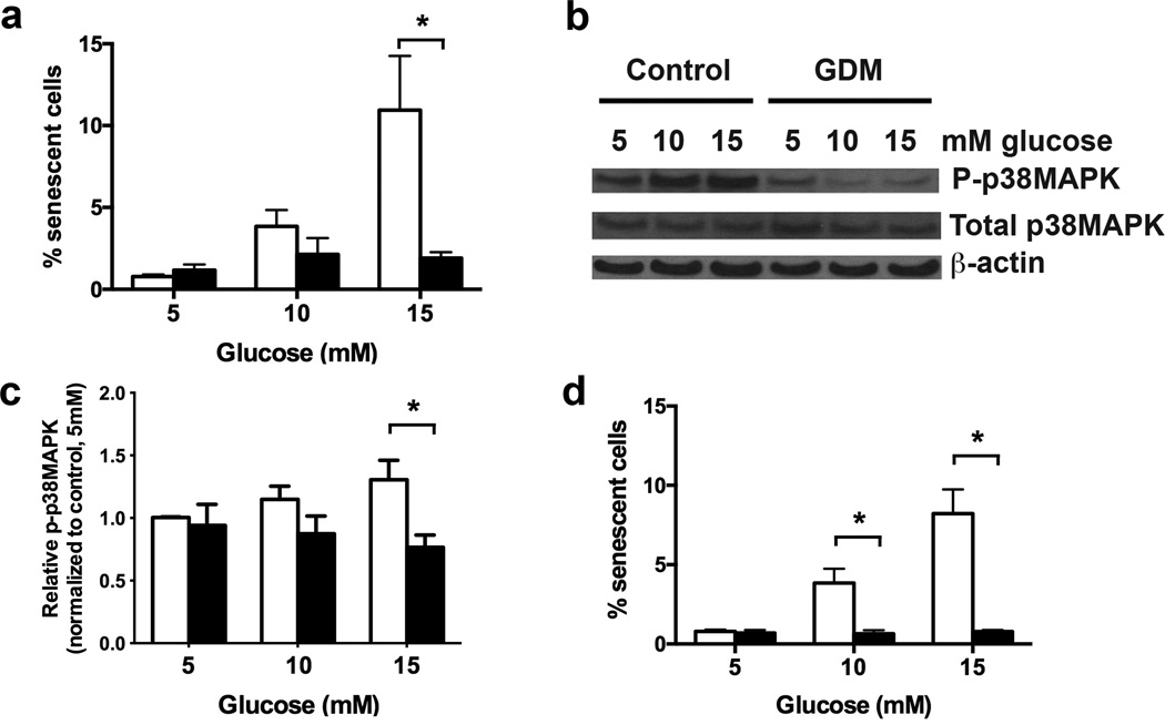
Figure 3. Hyperglycemic treatment of GDM ECFCs did not induce senescence or activate p38MAPK.
(a) Hyperglycemia-induced senescence. ECFCs were plated in EGM2 containing the indicated glucose concentration for 7 days. The percentage of SA-β-gal positive cells was calculated in 4 independent experiments. White bars indicate control ECFCs, and black bars indicated GDM ECFCs. Two-way ANOVA determined a significant increase in senescence by hyperglycemia, and with significantly less senescence in GDM ECFCs. *p<0.05 by Sidak’s multiple comparison. (b) Hyperglycemia induced p38MAPK phosphorylation in ECFCs. Western blotting was performed on lysates from control ECFCs treated with indicated glucose concentrations to detect phospho-p38MAPK and total p38MAPK. β-actin was used as a loading control. (c) Quantitation of band intensity from Western blots in panel (b) in 6 independent experiments. Bands were quantified in ImageJ. Phospho-p38MAPK (P-p38MAPK) levels were normalized to total p38MAPK. A significant increase in p38MAPK phosphorylation with higher glucose concentration was found by 2-way ANOVA. Decreased phospho-p38MAPK was found in GDM ECFCs as indicated (*p<0.05) by Sidak’s multiple comparisons. (d) Effect of p38MAPK inhibition on senescence. Control ECFCs were cultured in 5, 10, or 15 mmol/l glucose with or without 1 µM of the p38MAPK inhibitor, SB203580. White bars indicate control ECFCs, and black bars indicated GDM ECFCs. After 7 days, the percentage of SA-β-gal positive cells was quantified. Results are from 4 independent experiments. SB203580 signficantly decreased senescence, with *p<0.05 as indicated (two-way ANOVA followed by Sidak’s multiple comparisons).

