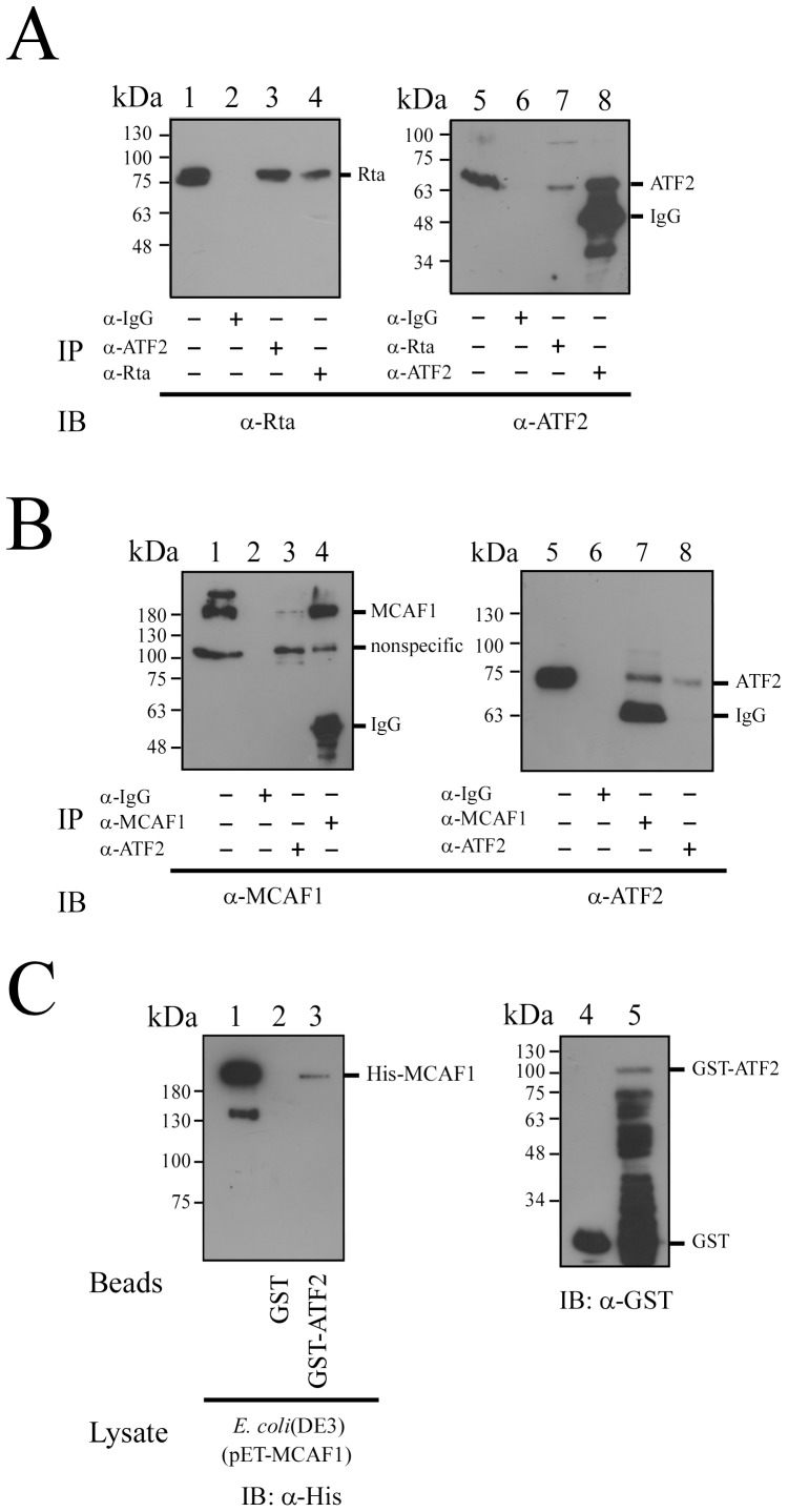
Figure 2. Interaction between Rta, MCAF1 and ATF2.
P3HR1 cells were treated with TPA and sodium butyrate for 24(A) Proteins in the lysate were immunoprecipitated with anti-ATF2 antibody (lanes 3, 8), anti-Rta antibody (lanes 4, 7) or anti-IgG antibody (lanes 2, 6). Immunoblot analysis was performed using anti-Rta (lanes 1–4) and anti-ATF2 antibodies (lanes 5–8). (B) Proteins in the lysate from P3HR1 cells were immunoprecipitated with anti-IgG antibody (lanes 2, 6), anti-ATF2 antibody (lanes 3, 8), or anti-MCAF1 antibody (lanes 4, 7). Immunoprecipitated proteins were detected by immunoblotting, using anti-MCAF1 antibody (lanes 1–4) or anti-ATF2 antibody (lanes 5–8). Lanes 1 and 5 in (A) and (B) were loaded with 1% of total protein from the cell lysate. (C) GST-ATF2 (lane 5) or GST (lane 4) was added to the lysate prepared from E. coli BL21(DE3)(pET-MCAF1) (lane 1). Proteins bound to GST-ATF2 were pulled down by glutathione-Sepharose beads, and analyzed by immunoblotting (IB) with anti-His antibody (lanes 1–3). Lane 1 was loaded with 1% of cell lysate. GST or GST-ATF2 bound to glutathione-Sepharose beads were analyzed by immunoblotting (IB) with anti-GST antibody (lanes 4, 5).

