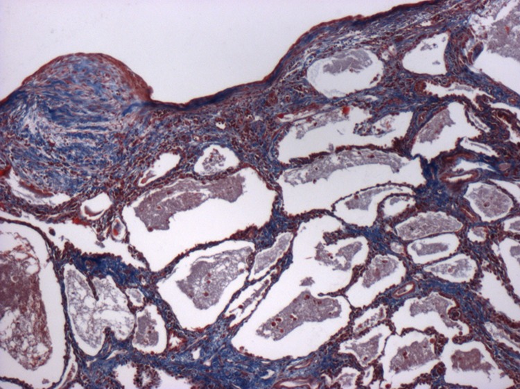
Figure 1. Low-power photomicrograph illustrating the characteristic appearances of usual interstitial pneumonia, the histological lesion of idiopathic pulmonary fibrosis (IPF).
The image shows fibroblastic foci overlying a region of micro-cystic honeycomb change. The sections have been stained with a Trichrome stain. This clearly highlights the extensive collagen (stained blue) and extracellular matrix deposition that occurs in IPF.

