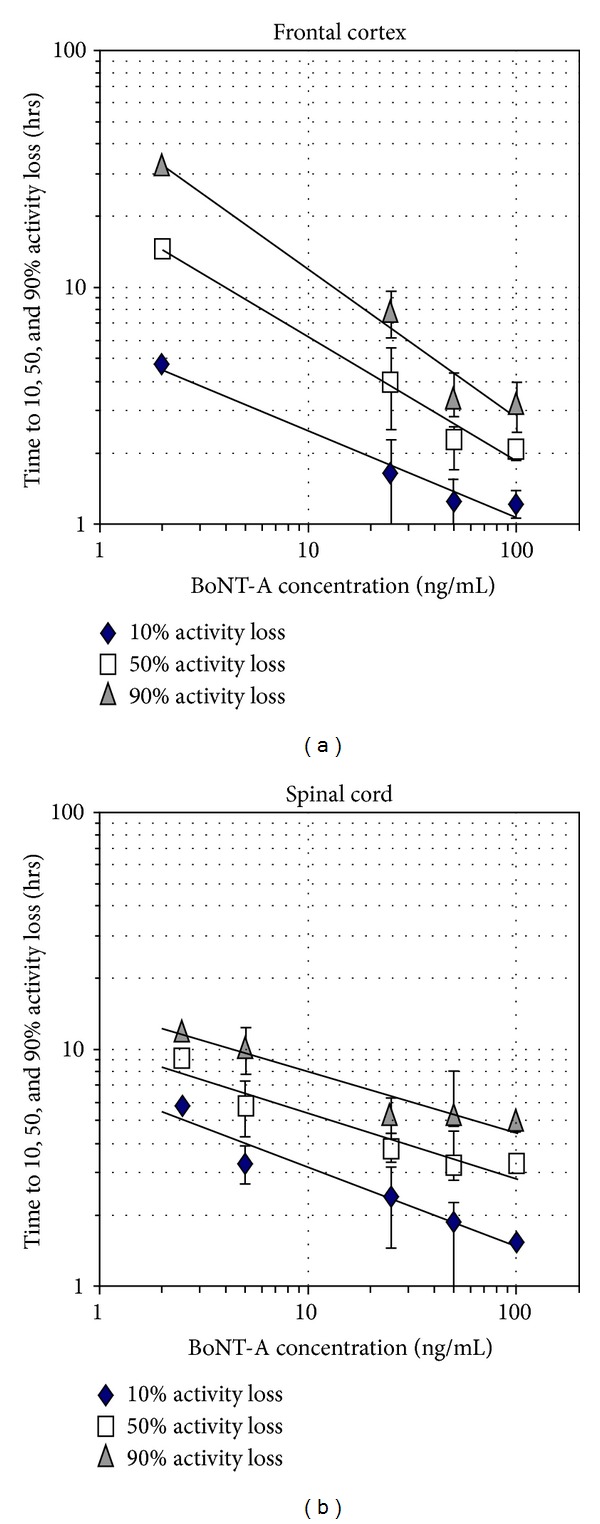
Figure 5.

Quantification of frontal cortex (a) and spinal cord (b) network responses to BoNT-A concentrations (x-axis; ng/mL). The time required to reach 10, 50, and 90 percent activity decreases is displayed on the y-axis. Both tissues generate approximate linear power function trend lines over the concentration range from 2 to 100 ng/mL (data points represent mean ± standard deviation).
