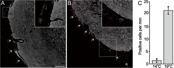
Figure 3.

Vascular endothelial growth factor (VEGF) expression in cardiac tissues of fish reared under normal and elevated temperature. Immunofluorescence staining of VEGF (red color) in cardiac tissues of fish reared for 56 days at control temperature (14°C, A) and elevated temperature (19°C, B). A: VEGF positive cells (arrows) are mainly located around already existing epicardial vasculature at 14°C. B: VEGF positive cells (arrows) are evenly distributed along the entire epicardium at 19°C. Panels A-B show one representative micrograph of three sections examined per fish from a total of three fish per temperature group, with one representative region per group shown at higher magnification (inset). The 400 μm scale bar in panel A applies to both panels in the figure. C: Average number of positive cells per mm epicardium ± SEM, calculated using a larger field of view (25× objective).
