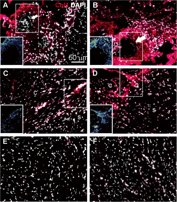
Figure 4.
Collagen I expression in cardiac tissues of fish reared at normal and elevated temperature. Immunofluorescence staining of collagen I (red color) and DAPI nuclear counterstain (white color) in cardiac tissues of fish reared for 56 days at 14°C (left panels, A, C, E) and 19°C (right panels, B, D, F). A: Collagen I is abundant in the epicardium and vasculature (arrow) at 14°C. B: Increased signal intensity is observed at 19°C. C: At 14°C collagen I is expressed in cell clusters in the compact (c) but not in the spongy (s) myocardium. D: At 19°C collagen I is strongly expressed in larger structures resembling connective tissue of the compact (c) but not in the spongy (s) myocardium. E-F: Cells in the spongy myocardium show weak staining at both temperatures. Fluorescence intensities are shown as LUT (Look-Up Table) images in panels A-D (inset). Panels show one representative micrograph of three sections examined per fish from a total of three fish per temperature group. The 50 μm scale bar in panel A applies to all panels in the figure.

