
Figure 3. Detection of reactive oxygen species in astrocytes in response to 24 hours of oxygen-glucose deprivation.
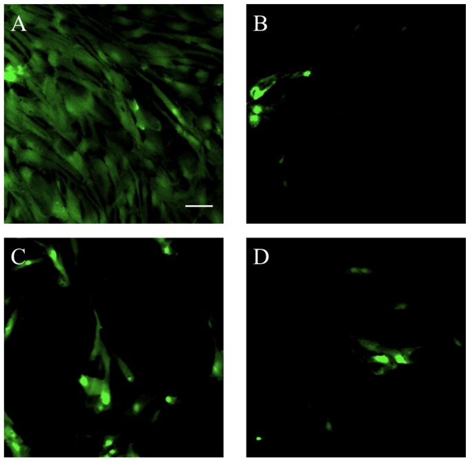
(A) Astrocytes that were exposed to OGD for 24 hours and treated with only serum-free, glucose-free media demonstrate a strong fluorescent signal indicating the production of ROS in astrocytes in response to OGD. Astrocytes that were treated with 10 ng/mL relaxin-2 (B), 10 ng/mL relaxin-3 (C) or 10 ng/mL R3/I5 (D) show a marked reduction in the production of ROS in response to OGD compared to untreated astrocytes (A). Scale bar = 50 µm.