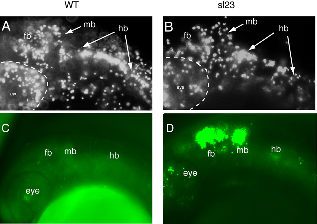
Figure 3. sl23 mutants show alterations in mitotic and apoptotic activities.
Anti-phospho-histone 3 staining of wild-type (WT) (A) and sl23 mutants (B) showed a decreased labeling throughout the head of the mutants, suggesting a reduction in the numbers of mitotically active cells. Acridine orange staining of dead cells in 24 hpf WT embryos (C) and sl23 mutant embryos (D) shows elevated cell death in the head of sl23 mutants, especially in the eye, forebrain and midbrain. Staining is much less in the hindbrain (hb).

