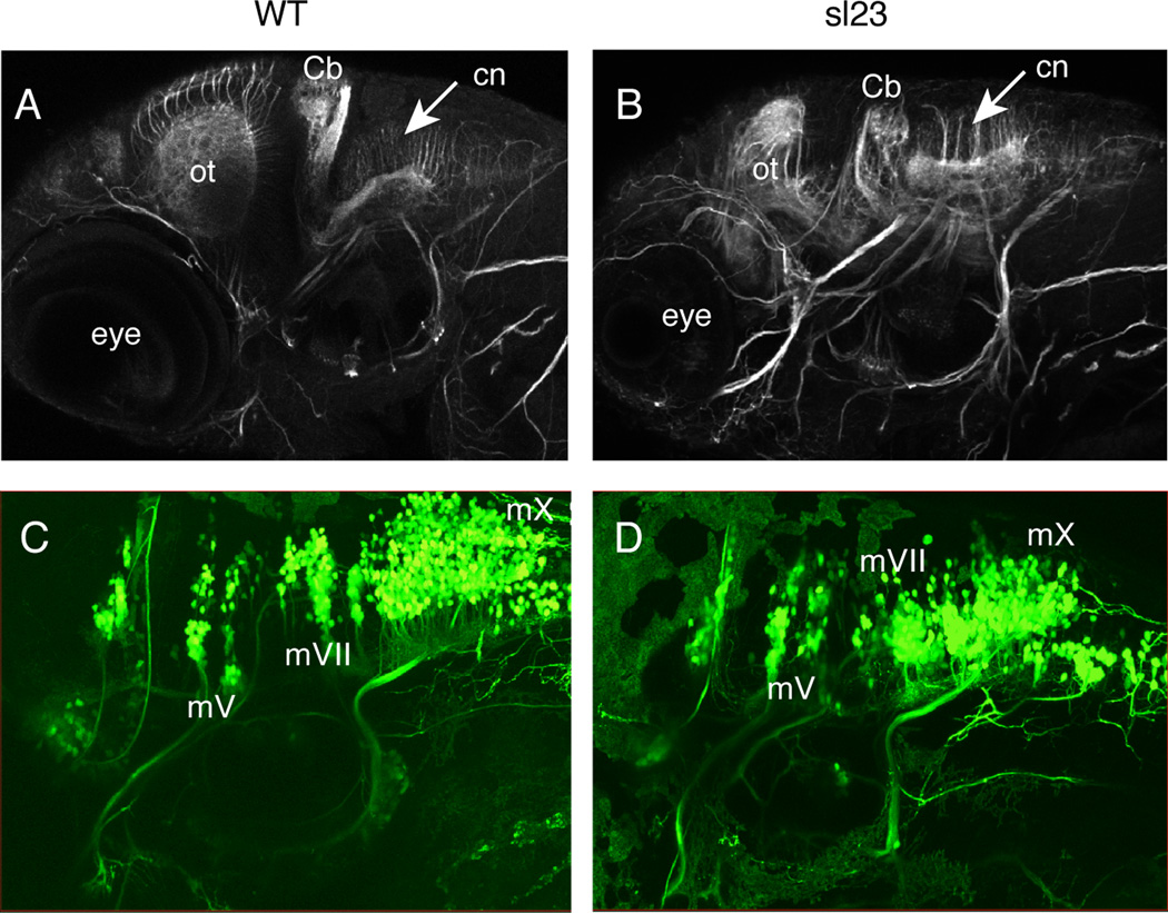
Figure 4. The hindbrain does not appear to be grossly affected in sl23 mutants.
Anti-acetylated tubulin immunostaining of 4 dpf WT (A) and sl23 mutant (B) larvae shows decreased size and structure of the optic tectum (ot) and cerebellum (Cb). Acetylated tubulin staining in the hindbrain of sl23 mutants appears to be almost normal, with the commissural neurons (cn) extending their axons dorsally, similar to the wildtype. There are no gross differences in the peripheral nerves between the sl23 mutants and WT. Confocal images of branchiomotor nerves at 4 dpf in WT (C) and sl23 mutant (D) larvae expressing the isl:gfp transgene show that the segmental organization of the motor nuclei in the hindbrain appears normal in sl23 compared to WT. The IXth and Xth motor nuclei in sl23 are a little compressed rostral to caudal, but their projections are normal. mV, Vth cranial motor nerve; mVII, VIIth cranial motor nerve; mX, Xth cranial motor nerve.

