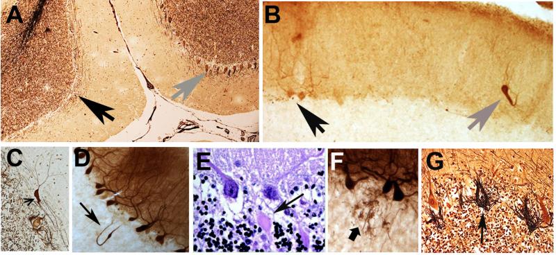
Figure 2.
A variety of structural microscopic changes have been described in the ET cerebellum on postmortem examination, and these seem to be centered on the Purkinje cell and surrounding neuronal populations (i.e., Basket cells). Cerebellar cortical sections in ET cases: A. Relatively normal Purkinje cell layer (gray arrow on right) near an area with segmental loss of Purkinje cells (black arrow on left). Bielschowsky stain. B. A heterotopic Purkinje cell (gray arrow) is adjacent to two normal Purkinje cells (black arrow). Calbindin-stained section. C. A swelling (arrow) of the Purkinje cell dendrite. Bielschowsky stain. D. A torpedo, thickened axon, and recurrent axonal collateral (arrow). Calbindin stain. E. Two torpedoes adjacent to their respective Purkinje cell soma. Luxol fast blue Hematoxylin and eosin stain. F. Sprouting of Purkinje cell axonal end processes in the upper granular layer. Calbindin stain. G. Hypertrophic Basket cell processes and an empty basket (arrow). Bielschowsky stain.

